Scientists recently boosted the resolution of magnetic resonance imaging (MRI) to 64 million times higher than normal. They used the technique to take captivating, high-definition images of a mouse brain, showing the organ like never before.
While the swirly, psychedelic images are that of a rodent's brain, the research team thinks humans could be next to undergo one of these newly enhanced brain scans. The technology could help doctors detect changes to the human brain that occur due to neurodegenerative diseases, such as Alzheimer's disease, as well as changes linked to healthy aging.
The mouse scan was shared as part of a new paper published April 17 in the journal PNAS (opens in new tab).
"It is something that is truly enabling," lead author G. Allan Johnson (opens in new tab), a distinguished professor of radiology at Duke University, said in a statement (opens in new tab). "We can start looking at neurodegenerative diseases in an entirely different way."
Related: First-ever scan of a dying human brain reveals life may actually 'flash before your eyes'
For four decades, Johnson, with the help of a revolving team of students and researchers from Duke University's Center for In Vivo Microscopy, has been working on improving MRI, which was invented by American physician Dr. Raymond Damadian (opens in new tab) 50 years ago.
MRI uses powerful magnets to generate magnetic fields, which cause the hydrogen atoms within water molecules in the body to align their "spins," or point in a specific direction. The machine then uses a pulse of radio waves to "flip" the atoms' spins. The atoms then fall back into alignment, and each flip generates a radio signal that can be detected by the MRI scanner and used to make an image.
To improve upon this technology, researchers created a souped-up MRI outfitted with a high-powered 9.4-tesla magnet. (For comparison, most MRIs are equipped with a 1.5- to 3-tesla magnet.) They also added gradient coils that are 100 times stronger than current models and are what create the images, as well as a high-speed computer that is as powerful as approximately 800 laptops, according to the statement.
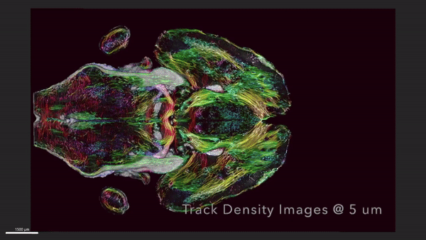
After scanning the mouse brain, the researchers sent tissue samples to be imaged using a technique called light sheet microscopy, which allowed them to label specific groups of cells in the brain that were then mapped onto the original MRI. These additional steps provided a colorful view of cells and circuits throughout the brain, according to the statement.
The researchers took one set of MRI images that captured how the mouse's brain-wide connectivity evolved with age. A second group of images showcased brilliantly colored brain connections that highlighted the deterioration of neural networks in a rodent model of Alzheimer's disease, according to the statement.
By studying mouse models of human diseases like Alzheimer's, researchers can better understand how these conditions emerge and progress in humans. The technique could also be useful for studying how the brain changes when mice are put on specific diets or given drugs in an effort to extend their life spans, Johnson said in the statement.
"The question is, is their brain still intact during this extended lifespan?" he said. "We have the capacity now to look at it. And as we do so, we can translate that directly into the human condition."


